Weizmann Institute AI Reconstructs Visuals From fMRI Scans With High Accuracy
Israeli researchers developed Brain-IT, an AI model that decodes brain activity to reconstruct images people view, requiring significantly less training data than previous methods.
Key points
- Brain-IT reconstructs viewed images from fMRI data with higher detail and speed than prior models.
- The system requires only one hour of subject-specific data to match results from forty-hour training sets.
- Researchers identified 128 functional brain regions to help the AI decode visual patterns.
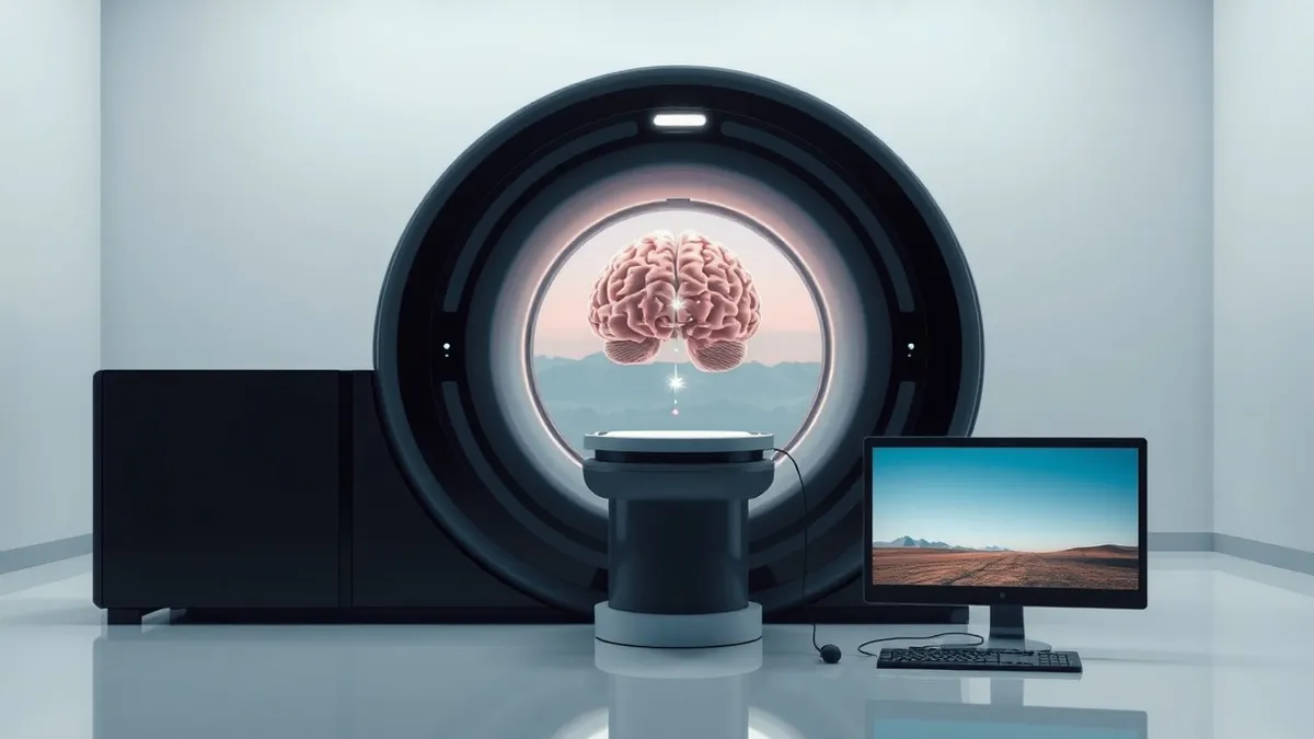
Researchers at the Weizmann Institute of Science in Israel have developed an artificial intelligence model capable of reconstructing visual images from functional magnetic resonance imaging brain scans. According to a report by CNET, the system, named Brain-IT, achieves startling accuracy in determining what individuals are seeing, effectively functioning as a form of mind-reading technology within laboratory settings.
The model was created by Professor Michal Irani and colleagues, who presented their findings at a scientific conference earlier this year. They documented the work on GitHub and in a published paper. The technology relies on pattern recognition to interpret data from fMRI scans, which track changes in blood flow and oxygen levels to measure neural activity. The AI then uses this physiological data to recreate the specific images the subject was viewing during the scan.
Technical Performance and Methodology
Brain-IT operates by identifying patterns in brain activity associated with specific visual stimuli. The researchers trained the model using thousands of brain scans sourced from the publicly available Natural Scenes Dataset. This dataset contained recordings from eight volunteers who viewed specific images while undergoing scanning procedures. The AI analyzed how different regions of the brain activated in response to these visual inputs, allowing it to map neural responses to visual content.
Professor Irani noted that previous AI models could translate brain activity into images but often failed to preserve basic features like composition and color. According to a statement from Irani, the new model outperforms existing methods in reconstructing both image content and fine details. The system also works in reverse, predicting what a brain scan would look like if a specific image were shown to a subject.
Significantly, Brain-IT requires substantially less training data than competing approaches. The researchers stated that the model needs only one hour of fMRI data from a new subject to achieve results comparable to other methods trained on forty hours of recording. This efficiency suggests the technology could be more practical for real-world applications where prolonged scanning is not feasible.
Potential Applications and Limitations
The primary potential applications for this technology lie in medical fields, particularly for helping individuals communicate who are unable to do so due to injury or disability. The researchers expressed hope that future advances could extend the technology’s capabilities to reading people’s dreams, though such applications remain speculative and distant. Currently, the work exists largely in lab settings and requires subjects to be inside an MRI machine.
The AI identifies 128 functional regions in the brain that activate when people think about specific things. For example, certain regions light up when a person looks at food, while others activate during the viewing of sports. Some of these regions were already known to neuroscientists, but the AI’s ability to correlate them with specific visual inputs enhances the reconstruction accuracy. The technology is limited to processing pictures and does not currently handle other sensory inputs or complex abstract thoughts.
What should you do?
Security and technology professionals should monitor developments in neural data processing and AI-driven biometric analysis. As these technologies move from laboratory experiments to potential commercial or medical products, the handling of sensitive neural data will become a critical privacy concern. Organizations involved in health tech or AI development should begin assessing the ethical and security implications of decoding brain activity.
What to do and how to stay safe: Weizmann Institute
- Monitor academic publications and conference proceedings for updates on Brain-IT and similar neural decoding technologies.
- Review data privacy policies to ensure they address the collection and storage of biometric and neural data, even if not currently in use.
- Assess current AI models for potential vulnerabilities related to pattern recognition and data inference techniques.
- Stay informed about regulatory developments concerning neuro-rights and the ethical use of brain-computer interfaces.
Step-by-step guide: Prompt Injection Warning Signs: How to Spot AI Manipulation
General security guidance from the Threat Times newsroom. It is not confirmed advice from the organisations named in this story.
Frequently asked questions
How does Brain-IT reconstruct images from brain scans?
The AI analyzes fMRI data that tracks blood flow and oxygen changes in the brain, using pattern recognition to match neural activity with visual content.
What is the training data requirement for Brain-IT?
The model requires only one hour of fMRI data from a new subject to match the accuracy of other methods that needed forty hours of recording.
Can this technology read dreams or thoughts outside of viewing images?
No, the technology currently only works with pictures viewed by subjects inside an MRI machine, though researchers hope to extend it to dreams in the future.